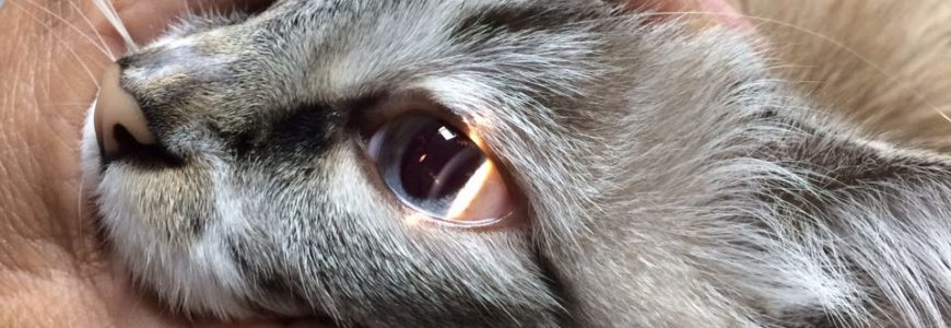
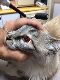
It consists of the use of a special portable microscope that allows us to perform a thorough examination of all the anatomical structures that surround and cover the eyeball, such as eyelids with some defect, eyelashes with abnormal birth or growth direction, fat producing glands, oil or water deficiency or atrophy that in many cases are the cause of the origin of the ophthalmologic problem.
This device in turn has some accessories, such as slit light which beams a light in the form of a vertical line that allows us to assess the thickness and depth of some structures (as well as possible injuries in them). It also has a cobalt blue light which serves as a contrast for some of the diagnosis dyes.