Corneal ulcers are very frequent affections, and they have associated incidence in some breeds, frequently due to the conformation of the face. Corneal ulcers tend to be a more common affliction in short snout dogs.
The corneal ulcer can be present as a slight superficial erosion that affects only the epithelium or first layer of the eye. An ulcer that does not go deeper into the second layer of the eye, called stroma, should heal within 7 to 8 days, unless complicated by infection due to lack of immediate appropriate treatment.
Another similar form of corneal ulcer, is the so-called indolent or torpid ulcer, obtaining its name from the difficulty in healing it presents. In this kind of ulcer, weeks or months can pass even under intense treatment with little progress. These ulcers are generally not very painful and have a rim that detaches easily becoming wider or moving from its original location. Their cause is the deficiency of the intra and extra-cellular ingredients necessary for the adhesion between the cells, so it is recommended to resort to other more specialized healing methods.
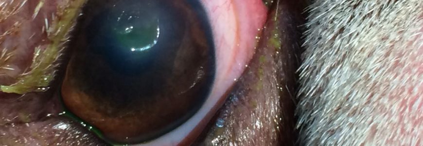
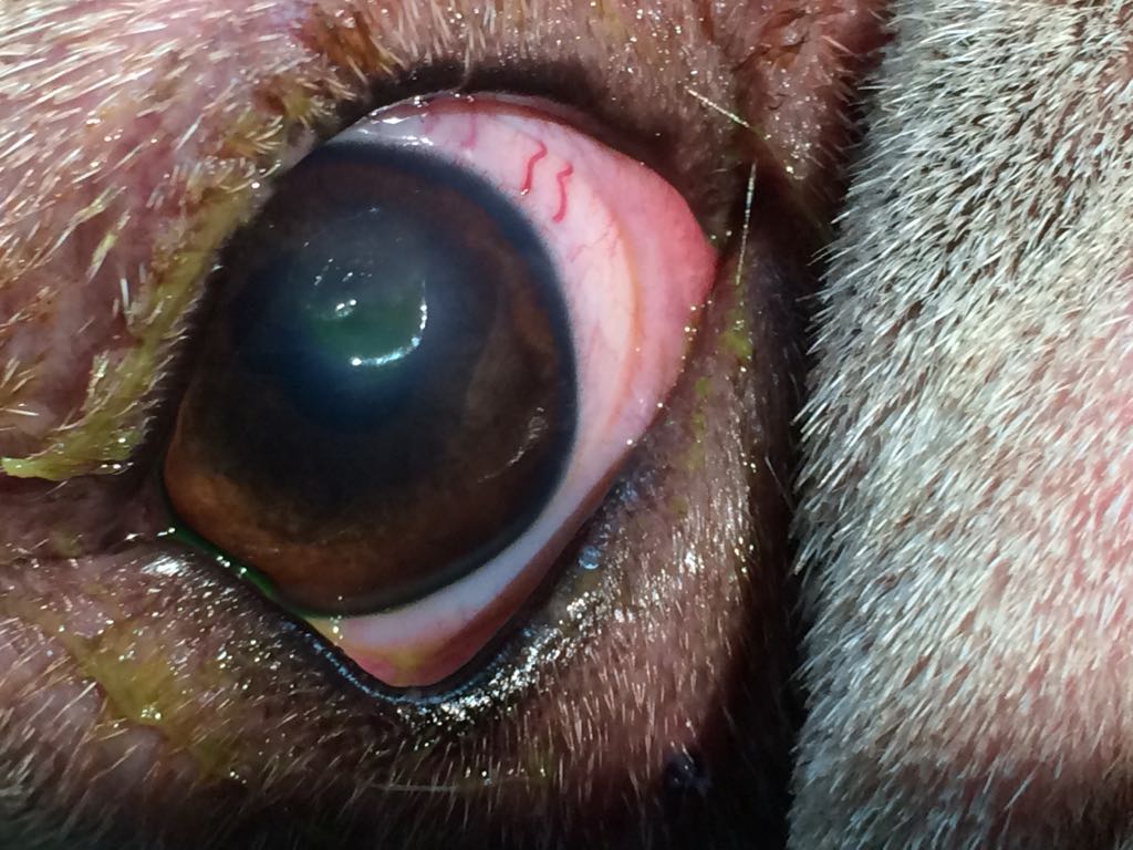
Stromal ulcers are those that go deeper into the second layer of the cornea. Those ulcers may or may not be infected and are more common in brachycephalic (short snout) dogs. The cause can be different factors, such as the rubbing of the cornea with hairs from some fold of the skin or by eyelids rolled inward, eyelashes directed towards the cornea, decrease or lack of lubrication by deficiency or absence of tears and also complications with viral bacterial infection or fungus. The different possible causes make it necessary to perform studies of secretions or of a cell sample, to facilitate diagnosis and prescribe the most effective treatment.
For the diagnosis of ulcers, a dye that stains when there is a lesion is applied. This can better be observed by directing a ray of cobalt blue light to the cornea.
This type of deeper ulcers are extremely painful so pet owners may notice spasms of the eyelid (when the eye is closed), excess tearing, and redness of the conjunctival membrane. When complicated with infection, it leads to uveitis, which is the internal inflammation of the entire eyeball where pus is sometimes observed internally. If not treated promptly and accurately, this could lead to blindness.
These ulcers usually have rapid progression that leads to descemetocele, i.e. the exposure of the last or third layer which is called the Descemet membrane. The Descemet membrane is extremely thin and delicate and is at risk of tearing or perforating at any time in these cases. Such tearing or perforation could lead to a further emptying of the internal liquid. In this case, the most indicated form of action is to prevent damage to the Descemet membrane, avoiding useless treatment, and proceeding to fix the ulcer surgically before it perforates the membrane. Case contrary, even when there would also be the need of a surgery in order to deal with the resulting problem, the outcome could be more uncertain.
Another type of ulcer is the liquefactive or collagenolytic ulcer. It is recognizable when the cornea starts to melt into a form of white gelatinous mucus until it perforates. The causes are many; it can be due to an infection by fungi or due to the presence of certain bacteria that produces destructive or toxic substances on the cornea. These type of ulcers cases are considered emergencies and hospitalization is required in order to give the pet intensive treatment and/or surgery, according to the particular case.